New technique can visualize your thoughts
Researchers have been trying to develop new tools in order to understand human mind. One important step have been already made, as a team of Dutch researchers from Donders Institute for Brain, Cognition and Behaviour in Nijmegen announced recently that a high-resolution fMRI, and computational modeling has allowed them to draw a subject’s experiences right out of the brain, reconstructing their thoughts. The study builds on years of research into how visual and cognitive information is represented in the brain.
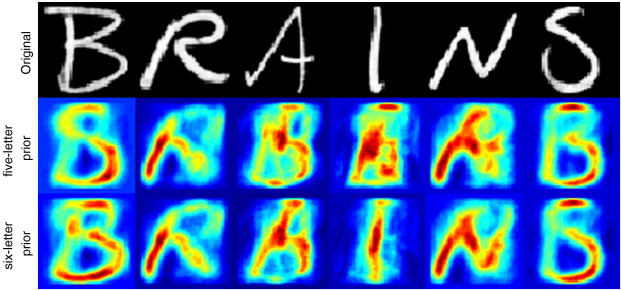 fMRI can identify the brain regions that use oxygen to do work during a particular task. Their fMRI data was reported in 2x2x2 millimeter units known as voxels, and the researchers showed subjects a particular letter, and their recordings of the resulting brain activity came in the form of these 3D pixels. They compiled a database of responses to different letters, creating a library of how the relevant brain areas visualize each different shape. Once this was done, recordings of a brain’s reaction to each letter could be checked against the database. However, they didn’t just get a basic letter identification, they actually reconstructed images of the letters themselves. Their algorithm translates brain voxels into image pixels, and it can learn how to do this more accurately as it compiles experience (machine learning algorithms). With an increase in resolution, they hope to upgrade from identifying letters to human faces. But they will still need to pass their data through the parsimony algorithm, pushing the results toward precollected standards. To create totally novel images from brain scans, to literally see what the subject sees like an intra-cranial camera, would require many large steps forward in our understanding of how the brain processes and conceptualizes visual data.
Even with the limitations, these scans could be a powerful tool and the potential is really exciting. It could even be used to prove someone’s innocence or guilt in court. So these results will be a major step forward for brain science.
More information can be found in the article that will soon be published in the journal NeuroImage: Linear reconstruction of perceived images from human brain activity (authors: Sanne Schoenmakers, Markus Barth, Tom Heskes, Marcel van Gerven).


